45 heart structure and labels
Heart Labeling Quiz: How Much You Know About Heart Labeling? Here is a Heart labeling quiz for you. The human heart is a vital organ for every human. The more healthy your heart is, the longer the chances you have of surviving, so you better take care of it. Take the following quiz to know how much you know about your heart. Questions and Answers. 1. Layers of the heart: Epicardium, myocardium, endocardium | Kenhub The epicardium is the outermost layer of the heart. It is actually the visceral layer of the serous pericardium, which adheres to the myocardium of the heart. Histologically, it is made of mesothelial cells, the same as the parietal pericardium. Below the mesothelial cells is a layer of adipose and connective tissue that binds the epicardium to ...
The structure of the heart - Structure and function of the heart ... It is located in the middle of the chest and slightly towards the left. The heart is a large muscular pump and is divided into two halves - the right-hand side and the left-hand side. The...
Heart structure and labels
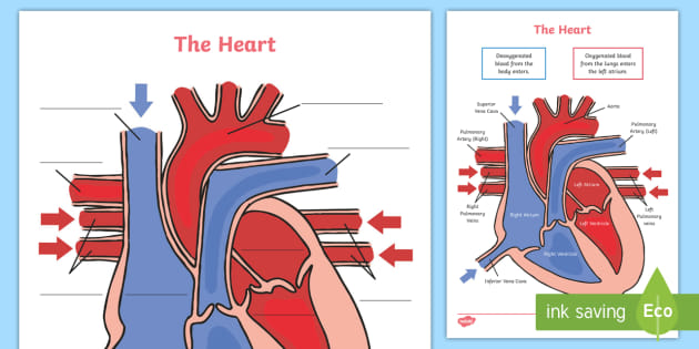
Labelling the heart — Science Learning Hub Blood transports oxygen and nutrients to the body. It is also involved in the removal of metabolic wastes. In this interactive, you can label parts of the human heart. Drag and drop the text labels onto the boxes next to the diagram. Selecting or hovering over a box will highlight each area in the diagram. Heart Anatomy Labeling Game About this Quiz This is an online quiz called Heart Anatomy Labeling Game There is a printable worksheet available for download here so you can take the quiz with pen and paper. Your Skills & Rank Total Points 0 Get started! Today's Rank -- 0 Today 's Points One of us! Game Points 19 You need to get 100% to score the 19 points available Actions Heart: Anatomy and Function What are the parts of the heart's anatomy? The parts of your heart are like the parts of a house. Your heart has: Walls. Chambers (rooms). Valves (doors). Blood vessels (plumbing). Electrical conduction system (electricity). Heart walls Your heart walls are the muscles that contract (squeeze) and relax to send blood throughout your body.
Heart structure and labels. Heart Labels - Printable or Custom Printed Stickers | Avery.com Use our free specialty shape label templates to easily personalize your heart labels online. Customize one of our free designs or upload your own graphics and then choose the printing option that works best for you. Order your blank heart labels or custom printed heart labels and stickers online and get free shipping on orders of $50 more. Heart Anatomy | Anatomy and Physiology - Lumen Learning The human heart consists of four chambers: The left side and the right side each have one atrium and one ventricle. Each of the upper chambers, the right atrium (plural = atria) and the left atrium, acts as a receiving chamber and contracts to push blood into the lower chambers, the right ventricle and the left ventricle. Human Heart - Anatomy, Functions and Facts about Heart The human heart is divided into four chambers, namely two ventricles and two atria. The ventricles are the chambers that pump blood and atrium are the chambers that receive the blood. Among which, the right atrium and ventricle make up the "right portion of the heart", and the left atrium and ventricle make up the "left portion of the heart." 5. Heart: illustrated anatomy - e-Anatomy - IMAIOS This interactive atlas of human heart anatomy is based on medical illustrations and cadaver photography. The user can show or hide the anatomical labels which provide a useful tool to create illustrations perfectly adapted for teaching. Anatomy of the heart: anatomical illustrations and structures, 3D model and photographs of dissection.
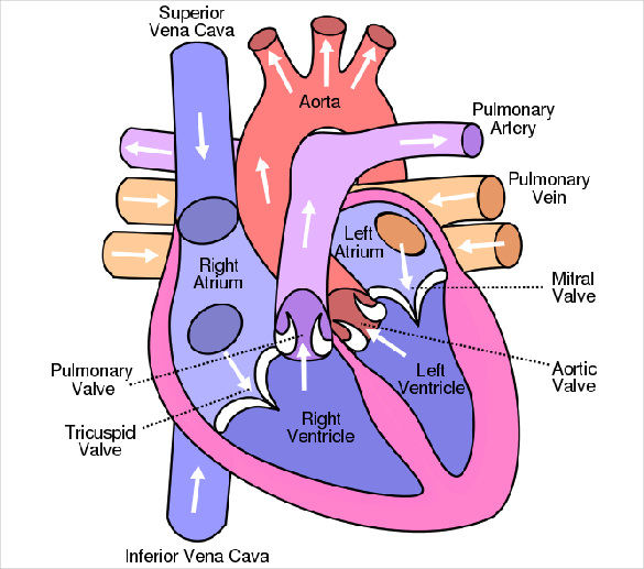
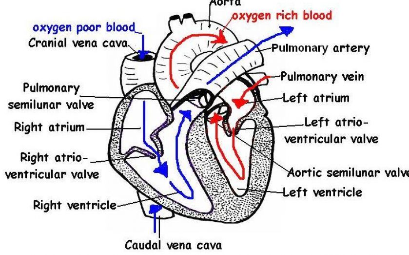
Label the heart — Science Learning Hub Label the heart Add to collection In this interactive, you can label parts of the human heart. Drag and drop the text labels onto the boxes next to the diagram. Selecting or hovering over a box will highlight each area in the diagram. Pulmonary vein Right atrium Semilunar valve Left ventricle Vena cava Right ventricle Pulmonary artery Aorta Dopamine - Wikipedia Structure. A dopamine molecule consists of a catechol structure (a benzene ring with two hydroxyl side groups) with one amine group attached via an ethyl chain. As such, dopamine is the simplest possible catecholamine, a family that also includes the neurotransmitters norepinephrine and epinephrine. The presence of a benzene ring with this amine attachment … Structure of the Heart | SEER Training The human heart is a four-chambered muscular organ, shaped and sized roughly like a man's closed fist with two-thirds of the mass to the left of midline. The heart is enclosed in a pericardial sac that is lined with the parietal layers of a serous membrane. The visceral layer of the serous membrane forms the epicardium. Layers of the Heart Wall A Diagram of the Heart and Its Functioning Explained in Detail The heart blood flow diagram (flowchart) given below will help you to understand the pathway of blood through the heart.Initial five points denotes impure or deoxygenated blood and the last five points denotes pure or oxygenated blood. 1.Different Parts of the Body ↓ 2.Major Veins ↓ 3.Right Atrium ↓ 4.Right Ventricle ↓ 5.Pulmonary Artery ↓ 6.Lungs
How to Draw the Internal Structure of the Heart (with Pictures) To draw the internal structure of a human heart, follow the steps below. Part 1 Finding a Diagram 1 To find a good diagram, go to Google Images, and type in "The Internal Structure of the Human Heart". Find an image that displays the entire heart, and click on it to enlarge it. 2 Find a piece of paper and something to draw with. Structure of the Heart | The Franklin Institute Structure of the Heart Although most people know that the human heart doesn't bear much resemblance to a heart drawn on a Valentine's Day card, the image can still be a useful way to learn and remember the parts of the heart. The heart consists of four chambers: two atria on the top and two ventricles on the bottom. Label the Heart Shows a picture of a heart with letters and blanks for practice with labeling the parts of the heart and tracing the flow of blood within the heart. How the Heart Works: Diagram, Anatomy, Blood Flow The heart is an amazing organ. It starts beating about 22 days after conception and continuously pumps oxygenated red blood cells and nutrient-rich blood and other compounds like platelets throughout your body to sustain the life of your organs.; Its pumping power also pushes blood through organs like the lungs to remove waste products like CO2.; This fist-sized powerhouse beats (expands and ...
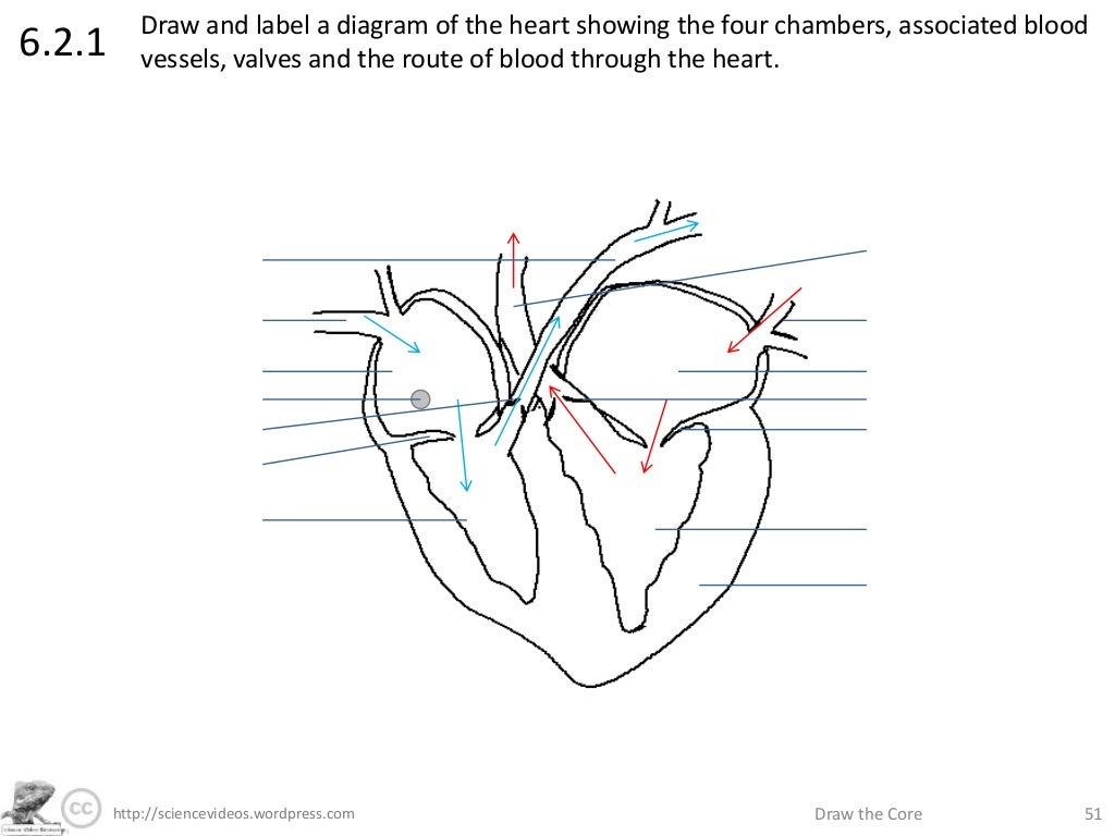
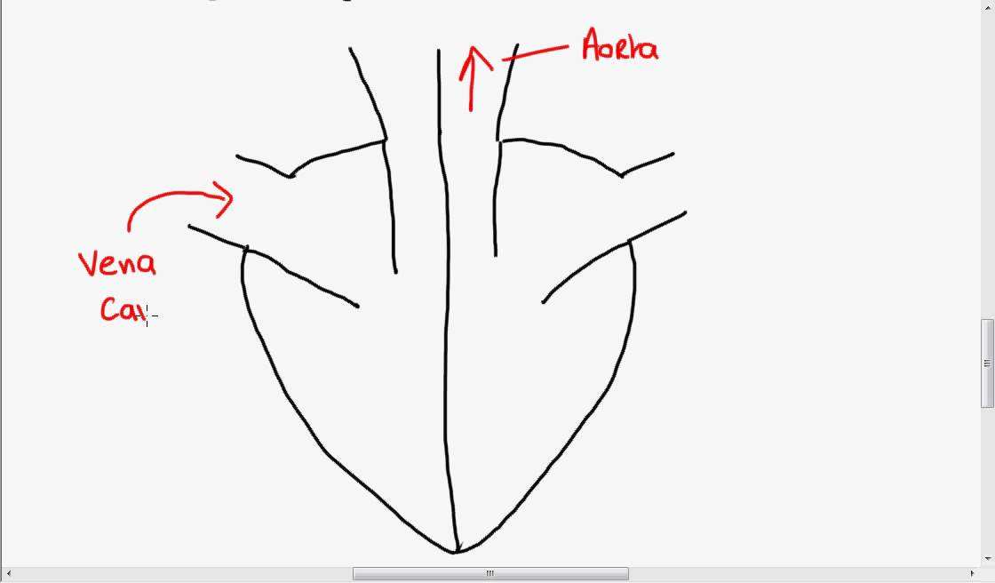
http://sciencevideos.wordpress.com Draw the Core 516.2.1
Human Heart Diagram Labeled | Science Trends List Of Heart Structures Heart Chambers Ventricles - The bottom two heart chambers. Atra - The upper two heart chambers. Wall Of The Heart Sinoatrial Node - A collection of tissue that releases electrical impulses and defines the rate of contraction for the heart. Atrioventricular Bundle - The fibers which transmit cardiac impulses.

The Heart Diagrams Labeled and Unlabeled
Human Heart (Anatomy): Diagram, Function, Chambers, Location in Body Chambers of the Heart The heart is a muscular organ about the size of a fist, located just behind and slightly left of the breastbone. The heart pumps blood through the network of arteries and...
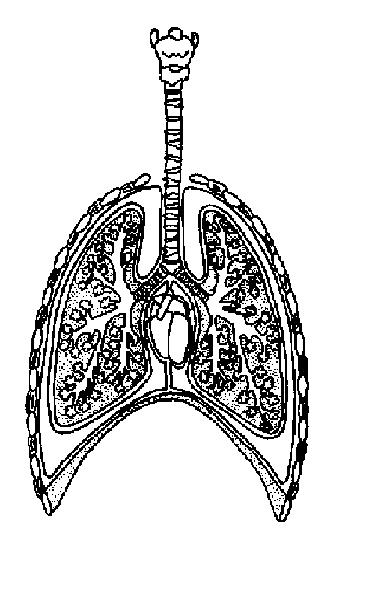
Respiratory System Worksheet - WikiEducator
Heart Anatomy: Labeled Diagram, Structures, Function, and Blood Flow There are 4 chambers, labeled 1-4 on the diagram below. To help simplify things, we can convert the heart into a square. We will then divide that square into 4 different boxes which will represent the 4 chambers of the heart. The boxes are numbered to correlate with the labeled chambers on the cartoon diagram.

Pin on Classical Conversations Science
Diagram of Human Heart and Blood Circulation in It A heart diagram labeled will provide plenty of information about the structure of your heart, including the wall of your heart. The wall of the heart has three different layers, such as the Myocardium, the Epicardium, and the Endocardium. Here's more about these three layers. Epicardium
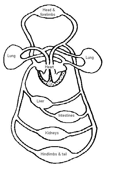
Circulatory System Worksheet - WikiEducator
Heart Anatomy: size, location, coverings and layers : Anatomy & Physiology Heart Anatomy. The heart is around the size of a fist and weighs between 250-350 grams (less than a pound). Enclosed within the mediastinum, the medial cavity of the thorax, the heart extends obliquely from the second rib to the fifth intercostal space. It rests on the superior surface of the diaphragm, lies posterior to the sternum and ...
Structure of the Heart - YouTube
Dapagliflozin - Wikipedia Dapagliflozin, sold under the brand names Farxiga (US) and Forxiga (EU) among others, is a medication used to treat type 2 diabetes. It is also used to treat adults with certain kinds of heart failure and chronic kidney disease.. Common side effects include hypoglycaemia (low blood sugar), urinary tract infections, genital infections, and volume depletion (reduced amount of …

Label Heart Structure | Medical Science Navigator
Label Heart Anatomy Diagram Printout - EnchantedLearning.com This cycle is then repeated. Every day, the heart pumps about 2,000 gallons (7,600 liters) of blood, beating about 100,000 times. Label the heart anatomy diagram below using the heart glossary. Note: On the diagram, the right side of the heart appears on the left side of the picture (and vice versa) because you are looking at the heart from the ...
Heart Diagram – 15+ Free Printable Word, Excel, EPS, PSD Template Download | Free & Premium ...
Heart Diagram with Labels and Detailed Explanation - BYJUS Diagram of Heart. The human heart is the most crucial organ of the human body. It pumps blood from the heart to different parts of the body and back to the heart. The most common heart attack symptoms or warning signs are chest pain, breathlessness, nausea, sweating etc. The diagram of heart is beneficial for Class 10 and 12 and is frequently ...

Eclectic Photography Project: Day 144 - strolling around Eureka, CA
Heart Blood Flow | Simple Anatomy Diagram, Cardiac Circulation ... - EZmed One of the first things you will notice if you look at the 12 steps is the pattern between the right and left side of the heart is similar. Step 1 and 6 involve a blood vessel, which makes sense as this is how blood enters and exits that side of the heart. Steps 2-5 involve a chamber, valve, chamber, and valve.
![heart labeling diagram | New Page 1 [jb004.k12.sd.us] | Photography | Pinterest | Heart anatomy ...](https://i.pinimg.com/originals/95/3a/5f/953a5f75ad2182832662829b56cf4684.jpg)
heart labeling diagram | New Page 1 [jb004.k12.sd.us] | Photography | Pinterest | Heart anatomy ...
The Anatomy of the Heart, Its Structures, and Functions The heart is the organ that helps supply blood and oxygen to all parts of the body. It is divided by a partition (or septum) into two halves. The halves are, in turn, divided into four chambers. The heart is situated within the chest cavity and surrounded by a fluid-filled sac called the pericardium. This amazing muscle produces electrical ...

Quia - 2015-01-12 Quiz - PLO JK - Heart Structure
Human Heart - Diagram and Anatomy of the Heart - Innerbody The heart is a muscular organ about the size of a closed fist that functions as the body's circulatory pump. It takes in deoxygenated blood through the veins and delivers it to the lungs for oxygenation before pumping it into the various arteries (which provide oxygen and nutrients to body tissues by transporting the blood throughout the body).
Heart Diagram Labelling Activity
Structure Of The Heart | A-Level Biology Revision Notes The heart is a hollow muscular organ that lies in the middle of the chest cavity. It is enclosed in the pericardium, which protects the heart and facilitates its pumping action. The heart is divided into four chambers: The two atria (auricles): these are the upper two chambers. They have thin walls which receive blood from veins.
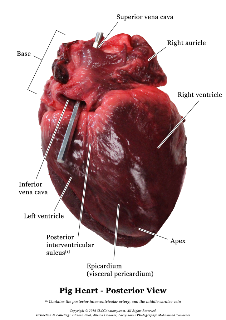
Pig Heart Dissection Photos - SLCC Anatomy
Heart: Anatomy and Function What are the parts of the heart's anatomy? The parts of your heart are like the parts of a house. Your heart has: Walls. Chambers (rooms). Valves (doors). Blood vessels (plumbing). Electrical conduction system (electricity). Heart walls Your heart walls are the muscles that contract (squeeze) and relax to send blood throughout your body.

Label Heart Structure Quiz
Heart Anatomy Labeling Game About this Quiz This is an online quiz called Heart Anatomy Labeling Game There is a printable worksheet available for download here so you can take the quiz with pen and paper. Your Skills & Rank Total Points 0 Get started! Today's Rank -- 0 Today 's Points One of us! Game Points 19 You need to get 100% to score the 19 points available Actions
Picture of the heart labeled
Labelling the heart — Science Learning Hub Blood transports oxygen and nutrients to the body. It is also involved in the removal of metabolic wastes. In this interactive, you can label parts of the human heart. Drag and drop the text labels onto the boxes next to the diagram. Selecting or hovering over a box will highlight each area in the diagram.
Post a Comment for "45 heart structure and labels"