40 picture of compound microscope with labels
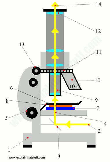
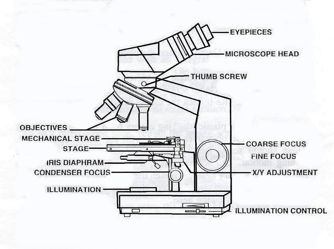
› cell-host-microbe › fulltextHost hepatic metabolism is modulated by gut microbiota ... May 26, 2022 · Identifying microbial metabolites modulating metabolism can be challenging. Le et al. use bioorthogonal chemistry to identify and trace the transfer of a unique bacterial sphingolipid to colon and liver tissue. Furthermore, bacterial sphingolipid synthesis by Bacteroides was determined to ameliorate hepatic lipid accumulation in a mouse model of hepatic steatosis. Compound Microscope - Diagram (Parts labelled), Principle and Uses See: Labeled Diagram showing differences between compound and simple microscope parts Structural Components The three structural components include 1. Head This is the upper part of the microscope that houses the optical parts 2. Arm This part connects the head with the base and provides stability to the microscope.
300+ Free Microscope & Laboratory Images - Pixabay Find your perfect microscope image. Free pictures to download and use in your next project. 189 37. analysis biochemistry. 335 71. analysis biochemistry. 334 96. microscope slide. 725 186.

Picture of compound microscope with labels
Compound Microscope High Resolution Stock Photography and Images - Alamy Find the perfect compound microscope stock photo. Huge collection, amazing choice, 100+ million high quality, affordable RF and RM images. ... Compound Microscope Stock Photos and Images (1,984) compound microscope isolated. Related searches: ... Method of illuminating compound microscope with gas lamp. Labels: C, ... Compound microscope Images, Stock Photos & Vectors - Shutterstock 3,117 compound microscope stock photos, vectors, and illustrations are available royalty-free. See compound microscope stock video clips Image type Orientation Sort by Popular Science College and University Biology Insects and Spiders Jobs/Professions microscope laboratory compound eye optical microscope scientist Next of 32 twitter.com › cityofcalgaryCity of Calgary (@cityofcalgary) | Twitter Aug 21, 2008 · Official City of Calgary local government Twitter account. Keep up with City news, services, programs, events and more. Not monitored 24/7.
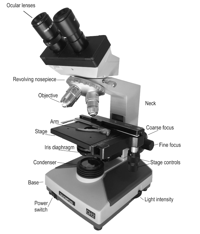
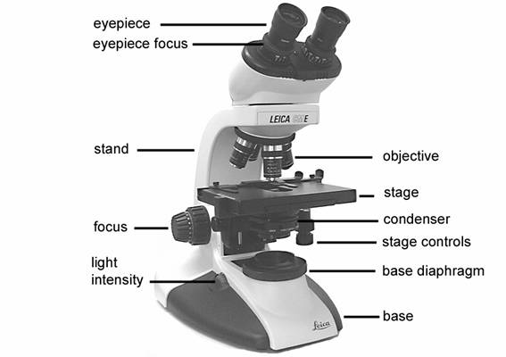
Picture of compound microscope with labels. A Study of the Microscope and its Functions With a Labeled Diagram To better understand the structure and function of a microscope, we need to take a look at the labeled microscope diagrams of the compound and electron microscope. These diagrams clearly explain the functioning of the microscopes along with their respective parts. ... The camera present within the microscope captures images to reveal the finer ... Compound Microscope Parts Made Easy A compound microscope employs a system of lenses and light to magnify the specimen. ... There are usually 3 or 4 objective lenses per microscope (the picture above has 3), and they usually have powers of 4x, 10x, 40x, and 100x respectively (one power per lens). ... Not labeled in the above diagram but you can see it as a lightly glowing circle ... Compound Microscope - Types, Parts, Diagram, Functions and Uses A compound microscope captures an inverted image of the specimen because every time the light passes through the lens, the image's direction is flipped. The image always ends up inverted from the original. So, if you move the sample to the left, it moves in the right direction. Image 18: A comparison image between a simple and compound microscope. Parts of a Compound Microscope - Labeled (with diagrams) Parts of a Compound Microscope - Labeled (with diagrams) A compound microscope is known as a high-power microscope that enables you to achieve a high level of magnification. Smaller specimens can be thoroughly viewed using a compound microscope. ... Picture 2: The basic parts of a compound microscope. image source : optimaxonline.com. Head/body
Compound Microscope Parts, Functions, and Labeled Diagram Compound Microscope Definitions for Labels. Eyepiece (ocular lens) with or without Pointer: The part that is looked through at the top of the compound microscope. Eyepieces typically have a magnification between 5x & 30x. Monocular or Binocular Head: Structural support that holds & connects the eyepieces to the objective lenses. Compound Microscope: Definition, Diagram, Parts, Uses, Working ... - BYJUS A microscope with a high resolution and uses two sets of lenses providing a 2-dimensional image of the sample. The term compound refers to the usage of more than one lens in the microscope. Also, the compound microscope is one of the types of optical microscopes. The other type of optical microscope is a simple microscope. Compound Microscope Parts - Labeled Diagram and their Functions - Rs ... Basically, compound microscopes generate magnified images through an aligned pair of the objective lens and the ocular lens. In contrast, "simple microscopes" have only one convex lens and function more like glass magnifiers. [In this figure] Two "antique" microscopes played significant roles in the history of biology. Microscope Types (with labeled diagrams) and Functions A compound microscope: Is used to view samples that are not visible to the naked eye Uses two types of lenses - Objective and ocular lenses Has a higher level of magnification - Typically up to 2000x Is used in hospitals and forensic labs by scientists, biologists and researchers to study micro organisms Compound microscope labeled diagram
Parts of the Microscope with Labeling (also Free Printouts) Microscopes are specially created to magnify the image of the subject being studied. This exercise is created to be used in homes and schools. the microscope layout, including the blank and answered versions are available as pdf downloads. Click to Download : Label the Parts of the Microscope (A4) PDF print version. Parts of a microscope with functions and labeled diagram Head - This is also known as the body. It carries the optical parts in the upper part of the microscope. Base - It acts as microscopes support. It also carries microscopic illuminators. Arms - This is the part connecting the base and to the head and the eyepiece tube to the base of the microscope. The Parts of a Microscope (Labeled) Printable - TeacherVision The Parts of a Microscope (Labeled) Printable. Download. Add to Favorites. Share. This diagram labels and explains the function of each part of a microscope. Use this printable as a handout or transparency to help prepare students for working with laboratory equipment. Microscope Parts and Functions Microscope Parts and Functions With Labeled Diagram and Functions How does a Compound Microscope Work?. Before exploring microscope parts and functions, you should probably understand that the compound light microscope is more complicated than just a microscope with more than one lens.. First, the purpose of a microscope is to magnify a small object or to magnify the fine details of a larger ...
How does a microscope work? - Explain that Stuff
recorder.butlercountyohio.org › search_records › subdivisionWelcome to Butler County Recorders Office Copy and paste this code into your website. Your Link Name
High Quality Labeled Diagram Of A Compound Microscope Photos - ClipArt Best - ClipArt Best
932 Compound Microscope Stock Photos - Dreamstime Browse 932 professional compound microscope stock photos available royalty-free. Microscope in blue science medical technology laboratory background. Compound microscope in blue science medical technology laboratory background. Typical animal cell Center 400x. The typical animal cell can be seen here.
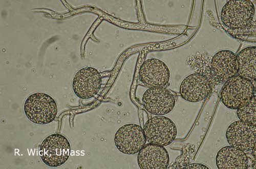
Greenhouse & Floriculture: Downy Mildews of Ornamental Plants | UMass Center for Agriculture ...
Parts of a Compound Microscope and Their Functions Compound microscope mechanical parts (Microscope Diagram: 2) include base or foot, pillar, arm, inclination joint, stage, clips, diaphragm, body tube, nose piece, coarse adjustment knob and fine adjustment knob. Base: It's the horseshoe-shaped base structure of microscope. All of the other components of the compound microscope are supported ...
Biology 130 Lab 1
happycamera.blog.jp › archives › 2020-082020年08月 : 写真で魅力発掘 ~暮らしのフォトダイアリー~ Powered by... Aug 31, 2020 · 中学生の息子と2人暮らしのシングルマザー。 シンプルで!元気に!楽しく!いられるように、試行錯誤しながらの日々です。 暮らしの話と、写真の撮り方のコツなどを発信。

35 Label Of Compound Microscope - Labels Information List
How to draw compound of Microscope easily - step by step - YouTube I will show you " How to draw compound of microscope easily - step by step "Please watch carefully and try this okay.Thanks for watching.....#microscopedrawi...

All Saints Online: Diagram for Labelling: Microscope
Compound Microscope Labeled Diagram - Quizlet QUESTION. The total magnification of a specimen being viewed with a 10X ocular lens and a 40X objective lens is. 15 answers. QUESTION. a mosquito beats its wings up and down 600 times per second, which you hear as a very annoying 600 Hz sound. if the air outside is 20 C, how far would a sound wave travel between wing beats. 2 answers.

Compound Microscope Clipart | Free Images at Clker.com - vector clip art online, royalty free ...
Labelled Diagram of Compound Microscope - Biology Discussion The below mentioned article provides a labelled diagram of compound microscope. Part # 1. The Stand: The stand is made up of a heavy foot which carries a curved inclinable limb or arm bearing the body tube. The foot is generally horse shoe-shaped structure (Fig. 2) which rests on table top or any other surface on which the microscope in kept.

34 Label A Compound Microscope - Labels Information List
Compound Microscope with labels Stock Vector | Adobe Stock Download Compound Microscope with labels Stock Vector and explore similar vectors at Adobe Stock. Adobe Stock Photos Illustrations Vectors Videos Audio Templates Free Premium Editorial Fonts
PRACTICAL BOOKLET - BIOLOGY4ISC
achieverstudent.comAchiever Student: Jul 28, 2020 · The best way to upload files is by using the “additional materials” box. Drop all the files you want your writer to use in processing your order.
3.5: Using the Compound Microscope - Biology LibreTexts
Microscope picture label Flashcards | Quizlet Start studying Microscope picture label. Learn vocabulary, terms, and more with flashcards, games, and other study tools.

Image result for olympus CX31 microscope labeled | Microscope, Olympus, Labels
prodottiplastici.roma.itSpi Driver Mpu9250 Jun 06, 2022 · Search: Mpu9250 Spi Driver. 00 P&P + £3 Last released Oct 11, 2017 MicroPython SPI driver for ILI934X based displays This is not needed when using a standalone AK8963 sensor An IMU (Inertial Measurement Unit) sensor is used to determine the motion, orientation, and heading of the robot Data is latched on the rising edge of SCLK Data is latched on the rising edge of SCLK.
:max_bytes(150000):strip_icc()/microscopestudy-58b978755f9b58af5c495ac7.png)
Learn About Microscopes With Fun, Free Printables
Microscope Diagram and Functions | Science fair projects ... - Pinterest A Study of the Microscope and its Functions With a Labeled Diagram To better understand the structure and function of a microscope, we need to take a look at the labeled microscope diagrams of the compound and electron microscope. These diagrams clearly explain the functioning of the microscopes along with their respective parts. M mooketsi

Euglena Acus 2 - BF microscope 1250x - YouTube
Compound Microscope- Definition, Labeled Diagram, Principle, Parts, Uses In order to ascertain the total magnification when viewing an image with a compound light microscope, take the power of the objective lens which is at 4x, 10x or 40x and multiply it by the power of the eyepiece which is typically 10x. Therefore, a 10x eyepiece used with a 40X objective lens will produce a magnification of 400X.
چگونه از میکروسکوپ نوری استفاده کنیم؟
Label the microscope — Science Learning Hub All microscopes share features in common. In this interactive, you can label the different parts of a microscope. Use this with the Microscope parts activity to help students identify and label the main parts of a microscope and then describe their functions. Drag and drop the text labels onto the microscope diagram.
Post a Comment for "40 picture of compound microscope with labels"