40 diagram of a cell with labels
Structure of Bacterial Cell (With Diagram) - Biology Discussion Cell wall: It is a tough and rigid structure of peptidoglycan with accessory specific materials (e.g. LPS, teichoic acid etc.) surrounding the bacterium like a shell and lies external to the cytoplasmic membrane. It is 10-25 nm in thickness. It gives shape to the cell. Nucleus: The single circular double-stranded chromosome is the bacterial genome. Venn diagram - Wikipedia A Venn diagram is constructed with a collection of simple closed curves drawn in a plane. According to Lewis, the "principle of these diagrams is that classes [or sets] be represented by regions in such relation to one another that all the possible logical relations of these classes can be indicated in the same diagram.That is, the diagram initially leaves room for any possible …
Converting Diagrams - The Biology Corner Open Google Draw and import the diagram. Then use "insert" to create text boxes where students can fill in the labels. Don't forget when assigning this to students on Google classroom to make a copy for each student. You can leave documents in an uneditable form and students can use an addon like "Kami" to annotate the document.
Diagram of a cell with labels
Animal Cells: Labelled Diagram, Definitions, and Structure - Research Tweet Animal Cells Organelles and Functions. A double layer that supports and protects the cell. Allows materials in and out. The control center of the cell. Nucleus contains majority of cell's the DNA. Popularly known as the "Powerhouse". Breaks down food to produce energy in the form of ATP. A Labeled Diagram of the Plant Cell and Functions of its Organelles ... A Labeled Diagram of the Plant Cell and Functions of its Organelles We are aware that all life stems from a single cell, and that the cell is the most basic unit of all living organisms. The cell being the smallest unit of life, is akin to a tiny room which houses several organs. Here, let's study the plant cell in detail... Interactive Cell Cycle - CELLS alive INTERPHASE. Gap 0. Gap 1. S Phase. Gap 2. MITOSIS . ^ Cell Cycle Overview Cell Cycle Mitosis > Meiosis > Get the Cell Division PowerPoints
Diagram of a cell with labels. PDF Human Cell Diagram, Parts, Pictures, Structure and Functions Diagram of the human cell illustrating the different parts of the cell. Cell Membrane The cell membraneis the outer coating of the cell and contains the cytoplasm, substances within it and the organelle. It is a double-layered membrane composed of proteins and lipids. How to draw an animal cell - labeled science diagram - YouTube Download a free printable outline of this video and draw along with us: you for watching. Please ... A Labeled Diagram of the Animal Cell and its Organelles One can observe the golgi apparatus in the labeled animal cell parts diagram. The golgi apparatus is situated near the cell nucleus and besides the stacked sacs, it also contains large number of vesicles. The main function of this golgi complex is to receive the proteins synthesized in the ER and transform it into more complex proteins. Feynman diagram - Wikipedia In theoretical physics, a Feynman diagram is a pictorial representation of the mathematical expressions describing the behavior and interaction of subatomic particles.The scheme is named after American physicist Richard Feynman, who introduced the diagrams in 1948.The interaction of subatomic particles can be complex and difficult to understand; Feynman diagrams give a …
Labeling a Cell Diagram | Quizlet Cell Wall This gives shape and support to the plant cell. It surrounds the cell and protects the other parts of the cell. Chloroplasts This is where the plant cell's chlorophyll is stored. This is what the plant uses to make its own food (photosynthesis). This is also what makes plant cells have a green-like color. Plant cells Are circular in shape Labeled Plant Cell With Diagrams | Science Trends The parts of a plant cell include the cell wall, the cell membrane, the cytoskeleton or cytoplasm, the nucleus, the Golgi body, the mitochondria, the peroxisome's, the vacuoles, ribosomes, and the endoplasmic reticulum. Parts Of A Plant Cell The Cell Wall Let's start from the outside and work our way inwards. Plant Cell Anatomy - Enchanted Learning The cell membrane is semipermeable, allowing some substances to pass into the cell and blocking others. Cell wall A thick, rigid membrane that surrounds a plant cell. This layer of cellulose fiber gives the cell most of its support and structure. The cell wall also bonds with other cell walls to form the structure of the plant. Cell Organelles- Definition, Structure, Functions, Diagram In a plant cell, the cell wall is made up of cellulose, hemicellulose, and proteins while in a fungal cell, it is composed of chitin. A cell wall is multilayered with a middle lamina, a primary cell wall, and a secondary cell wall. The middle lamina contains polysaccharides that provide adhesion and allow binding of the cells to one another.
Skin Diagram with Detailed Illustrations and Clear Labels Explore Skin Diagram with BYJU’S. Diagram of the skin is illustrated in detail with neat and clear labelling. Also available for free download. Login. Study Materials. ... Human Cell Structure: Types Of Microbes: Biome Meaning: What Is A Neuron: 1 Comment. Neeraj Shukla September 23, 2021 at 1:28 pm. Up board English medium. Reply. ^ Cell Cycle Overview Cell Cycle Mitosis > Meiosis > INTERPHASE. Gap 0. Gap 1. S Phase. Gap 2. MITOSIS . ^ Cell Cycle Overview Cell Cycle Mitosis > Meiosis > Get the Cell Division PowerPoints Venn diagram - Wikipedia A Venn diagram is a widely used diagram style that shows the logical relation between sets, popularized by John Venn (1834–1923) in the 1880s. The diagrams are used to teach elementary set theory , and to illustrate simple set relationships in probability , logic , statistics , linguistics and computer science . cell diagram to label Plant Cell Diagram And Label Simple - Cell Diagram diagram.oyajino.com. Nucleus And Ribosomes (article) | Khan Academy . nucleus ribosomes diagram structure labeled cell biology eukaryotic cells which khan. 2.3.1 Draw And Label A Diagram Of The Ultrastructure Of A Liver Cell As
Epithelia: The Histology Guide
Draw a diagram of a plant cell and label at least eight ... Hint: Plant cell has cell wall whereas cell wall is absent in an animal cell. The chloroplast is present only in plant cells. Plant cells contain large ...
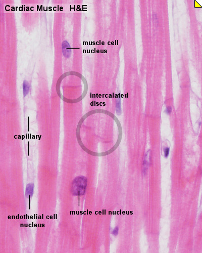
Cardiac Muscle Histology - Embryology
Cell Diagrams with Labelling Activity - Learnful Cell Diagrams with Labelling Activity ... I've created two interactive diagrams for an upcoming open textbook for high-school level biology. The cell structure ...
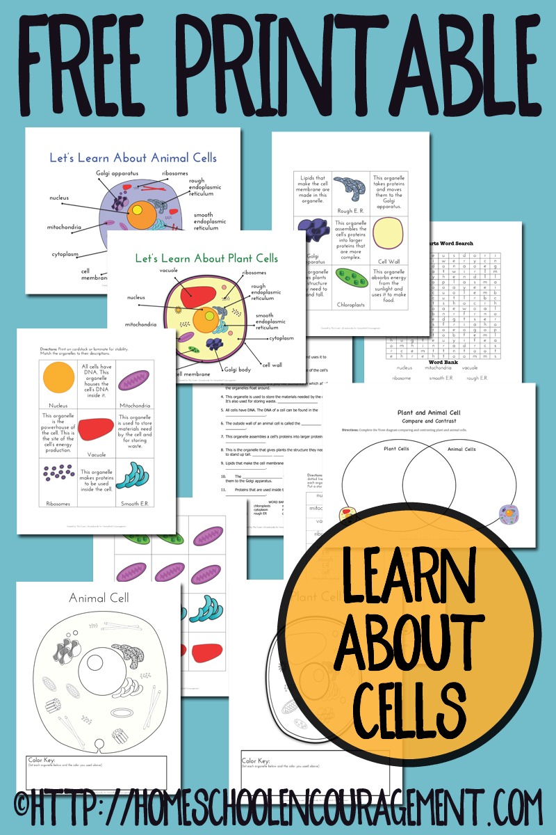
FREE Plant and Animal Cell Printables | Free Homeschool Deals
Plant Cell Diagram | Science Trends A plant cell diagram, like the one above, shows each part of the plant cell including the chloroplast, cell wall, plasma membrane, nucleus, mitochondria, ribosomes, etc.A plant cell diagram is a great way to learn the different components of the cell for your upcoming exam. Plants are able to do something animals can't: photosynthesize.Plant cells are able to do this because plant cells have ...
/plant_anaphase-56a09b0d3df78cafdaa32db4.jpg)
Daughter Chromosome
Structure of Cell: Definition, Types, Diagram, Functions - Embibe Structure of Cell: Cell is the basic functional unit that makes up all living organisms.All organisms, including ourselves, start life as a single cell called the egg. Cells are small microscopic units that perform all essential functions of life and are capable of independent existence.
.jpg)
Biology 101, Bledsoe
How to Create Venn Diagram in Excel – Free Template Download Replace the default values with the custom labels you previously designed. Right-click on any data label and choose “Format Data Labels.” Once the task pane pops up, do the following: Go to the Label Options tab. Click “Value From Cells.” Highlight the corresponding cell from column Label (H2 for Coca-Cola, H3 for Pepsi, and H4 for Dr ...
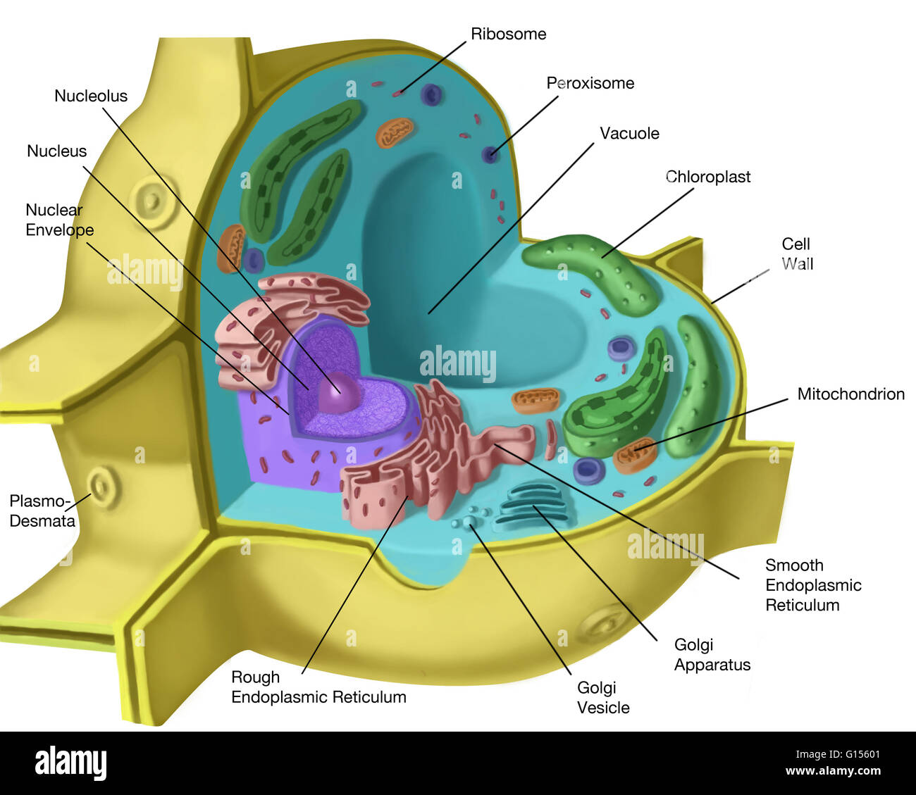
Plant cell structure. Artwork of a sectioned plant cell. The features Stock Photo - Alamy
Skin Diagram with Detailed Illustrations and Clear Labels - BYJUS Skin Diagram The largest organ in the human body is the skin, covering a total area of about 1.8 square meters. The skin is tasked with protecting our body from external elements as well as microbes.

Inspired Class: Create 3D Animal Cells with Play Doh
Chord diagram – from Data to Viz A chord diagram represents flows or connections between several entities (called nodes).Each entity is represented by a fragment on the outer part of the circular layout.Then, arcs are drawn between each entities. The size of the arc is proportional to the importance of the flow. Here is an example displaying the number of people migrating from one country to another.
.PNG)
Electrical Circuits - Presentation Physics
Cell Diagram To Label Teaching Resources | Teachers Pay Teachers On page 1 of this worksheet there is a photo sample of an onion root showing all 6 steps of the cell cycle.On page 2 of this worksheet the 6 steps are described with a diagram and students are instructed to find and label these stages on the onion root.
Post a Comment for "40 diagram of a cell with labels"